From EPpedia
(Difference between pages)
Jump to navigation
Jump to search
|
|
| Line 1: |
Line 1: |
| {{DevelopmentPhase}}
| | [[File:After_tabatabaei_and_asirvatham.svg|thumb]] |
| | | [[File:Yamashina_rvot_RFCA_CARTO.svg|thumb]] |
| {|style="border-spacing:8px;margin:0px 0x;align:center;" width="100%"
| | [[File:Jadonath_RVOT_localisation_algorythm.svg|thumb]] |
| | style="width:34%;border:1px solid #E2ACB1;background-color:#FFF5F5;vertical-align:top;color:#000"|
| | [[File:Ablation.svg|thumb|Schematic demonstrating the orientation of the right ventricular outflow tract (RVOT) in the chest cavity. AO = aorta.]] |
| {|width="100%" cellpadding="2" cellspacing="5" style="vertical-align:top;background-color:#FFF5F5"
| | [[File:Stevenson.svg|thumb|ECG types of VT and most common causes are shown with characteristic |
| ! <h2 style="margin:0;background-color:#D1DAEB;font-size:120%;font-weight:bold;border:1px solid #a3bfb1;text-align:center;color:#000;padding:0.2em 0.4em;">Devices</h2>
| | ECG features of selected VTs. LBBB indicates left bundle-branch block; LVOT, LV outflow tract; RBBB, right bundle-branch block; L, left; and R, right.]] |
| |-
| | <cite>test</cite> |
| |[[Image:Course.jpg|140px|center]]
| | <biblio> |
| |-valign="top"
| | #test ate |
| |
| | </biblio> |
| #[[Pacemakers]]
| |
| ##[[Pacemaker modi]]
| |
| ##[[Pacemaker programs and settings]]
| |
| ##[[Pacemaker troubleshooting]]
| |
| ##[[Pacemaker complications]]
| |
| #[[ICD]]
| |
| | |
| |}<!-- Start of right-column -->
| |
| | style="width:33%;border:1px solid #E2ACB1;background-color:#FFF5F5;vertical-align:top"|
| |
| {|width="100%" cellpadding="2" cellspacing="5" style="vertical-align:top;background-color:#FFF5F5"
| |
| !
| |
| | |
| <h2 style="margin:0;background-color:#D1DAEB;font-size:120%;font-weight:bold;border:1px solid #a3bfb1;text-align:center;color:#000;padding:0.2em 0.4em;">Electrophysiology</h2>
| |
| |-
| |
| |[[Image:book.jpg|140px|center]]
| |
| |-
| |
| |
| |
| *[[Basics]]
| |
| |}<!-- Start of right-column -->
| |
| |style="width:34%;border:1px solid #E2ACB1;background-color:#FFF5F5;vertical-align:top;color:#000"|
| |
| {|width="100%" cellpadding="2" cellspacing="5" style="vertical-align:top;background-color:#FFF5F5"
| |
| !
| |
| | |
| <h2 style="margin:0;background-color:#D1DAEB;font-size:120%;font-weight:bold;border:1px solid #a3bfb1;text-align:center;color:#000;padding:0.2em 0.4em;">Cases</h2>
| |
| |-
| |
| |[[Image:cases.jpg|140px|center]]
| |
| |-
| |
| |Casus:
| |
| *[[EGM tracings]]
| |
| |}
| |
| |}
| |
| | |
| | |
| | |
| | |
| [[PM and ICD manuals]]
| |
Revision as of 09:39, 12 December 2011
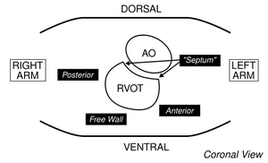
Schematic demonstrating the orientation of the right ventricular outflow tract (RVOT) in the chest cavity. AO = aorta.
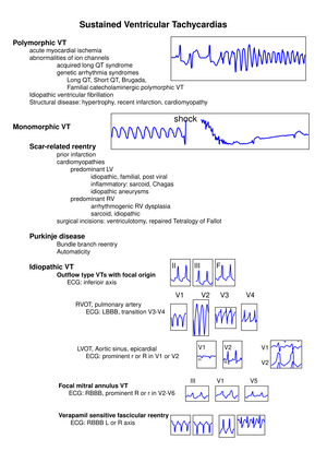
ECG types of VT and most common causes are shown with characteristic ECG features of selected VTs. LBBB indicates left bundle-branch block; LVOT, LV outflow tract; RBBB, right bundle-branch block; L, left; and R, right.
[1]
- [test]


