RVOT / LVOT tachycardia: Difference between revisions

From EPpedia
Jump to navigation Jump to search
No edit summary
No edit summary
Line 7: Line 7:
<cite>test</cite>
<cite>test</cite>
<biblio>
<biblio>
#test pmid=100
#test pmid=1000
</biblio>
</biblio>

Revision as of 13:25, 14 December 2011

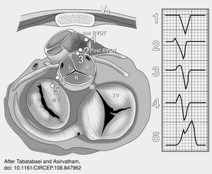
After tabatabaei and asirvatham.svg
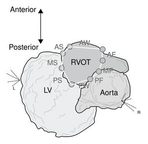
Yamashina rvot RFCA CARTO.svg
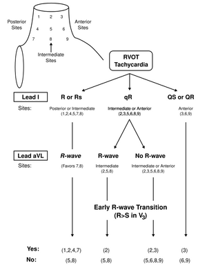
Jadonath RVOT localisation algorythm.svg
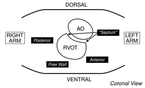
Schematic demonstrating the orientation of the right ventricular outflow tract (RVOT) in the chest cavity. AO = aorta.
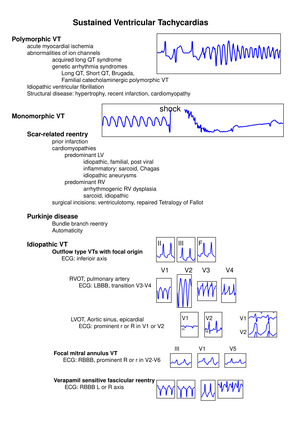
ECG types of VT and most common causes are shown with characteristic ECG features of selected VTs. LBBB indicates left bundle-branch block; LVOT, LV outflow tract; RBBB, right bundle-branch block; L, left; and R, right.

[1]

  1. Wootton JC, Taylor JG, Jackson AA, Chambers GK, and Fincham JR. The amino acid sequence of Neurospora NADP-specific glutamate dehydrogenase. The tryptic peptides. Biochem J. 1975 Sep;149(3):739-48. DOI:10.1042/bj1490739 | PubMed ID:1000 | HubMed [test]